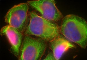
 Johns Hopkins researchers have used a small synthetic molecule to stimulate cells to move and change shape, bypassing the cells’ usual way of sensing and responding to their environment; this represents a new tool for studying cell movement: a phenomenon needed to understand cancer.
Many types of human cells move, including fibroblasts, which patrol the skin and make repairs; immune cells, which rush to the site of infections; nerve cells, which travel great distances during development and tumour cells that break off and migrate to a new part of the body.
In the natural process for stimulating cell movement, signalling proteins bind to receptor molecules on the surface of the cell, setting off a complex chain reaction that ultimately propels it in a certain direction. Unfortunately it is extremely difficult to study this process directly. A further difficulty is that direction of cell movement is controlled by differential signal concentration on either side of the cell. Replicating this process is not a trivial thing to do, because the cells are incredibly small.
In the study, the scientists employed a small molecule able to get in-between the molecules of the cell membrane; it binds to two slightly modified proteins, which then trigger the release of the critical protein, beginning the chain reaction that eventually leads to movement. In order to guide a cell in a specific direction, the researchers built a chip with tiny liquid-dispensing channels. When these channels are loaded with a solution containing the synthetic molecule and placed on the surface of human cells they could stimulate one side of a cell more than the other. The cells responded very dramatically, moved in the direction specified, and changed their shapes.